
2013 - 5 Issue

Case report

The Relations of Morphological and Functional Changes in Children with Retinal Dystrophy Disease

Full Text links

Summary

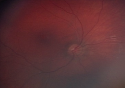
PURPOSE: To characterize the correlation between functional and morphological changes in the retina in the retinal dystrophies in children.

METHODS: In the group of six patients with selected types of retinal dystrophies was analysed the morphological findings obtained by the Optical coherence tomography (OCT) and their correlation with the electrophysiological findings.

RESULTS: Typical morphological retinal changes visualised by OCT were confirmed in all examined patients and were in correlation with progressive loss of visual function (decrease of visual acuity, constriction of visual field or scotomas in visual field, colour vision defect, nyctalopia) and abnormal values of the electrophysiological findings. CONCLUSIONS: Optical coherence tomography and electrophysiological methods are essential in approaching patient with tapetoretinal dystrophies. Correlation of these findings enables us to make diagnose easier, to understand better the dynamic of the morfological and functional changes in these patients. It can also be implicated as prognostic indicators for visual progression in patients with retinal dystrophy and also in prevention by means of genetic methods.

References

  1. 1. Alió JL, Plaza-Puche AB, Piñero DP, Javaloy J, Ayala MJ.: J Cataract Refract Surg, 2011; 37:1605–14.
  2. Auffarth GU, Wilcox M, Sims JCR, McCabe C, Wesendahl TA, Apple DJ.: Analysis of 100 explanted one-piece and three-piece silicone intraocular lenses. Ophthalmology, 1995; 102:1144–1150.
  3. Connors R III, Boseman P III, Olson RJ.: Accuracy and reproducibility of biometry using partial coherence interferometry. J Cataract Refract Surg, 2002; 28:235–238.
  4. Erie JC, Bandhauer MH, McLaren JW.: Analysis of postoperative glare and intraocular lens design. J Cataract Refract Surg, 2001; 27:614–621.
  5. Farbowitz MA, Zabriskie NA, Crandall AS, Olson RJ, Miller KM.: Visual complaints associated with the AcrySof acrylic intraocular lens. J Cataract Refract Surg, 2000; 26:1339–1345.
  6. Franchini A, Zamma Gallarati B, Vaccari E.: Computerized analysis of the effects of intraocular lens edge design on the quality of vision in pseudophakic patients. J Cataract Refract Surg, 2003; 29:342–347.
  7. Holladay JT, Lang A, Portney V.: Analysis of edge glare phenomena in intraocular lens edge designs. J Cataract Refract Surg, 1999; 25:748–752.
  8. Kuchynka, P.: Čočka in Kuchynka, P.: Oční lékařství, Grada Publishing, a.s. 2007, s. 372-426.
  9. Leaming DV.: Practice styles and preferences of ASCRS members - 2003 survey. J Cataract Refract Surg, 2004; 30:892–900.
  10. Mamalis N.: Complications of foldable intraocular lenses requiring explantation or secondary intervention - 1998 survey. J Cataract Refract Surg, 2000; 26:766–772.
  11. Mamalis N, Spencer TS.: Complications of foldable intraocular lenses requiring explantation or secondary intervention - 2000 survey update. J Cataract Refract Surg, 2001; 27:1310– 1317.
  12. Mamalis N.: Complications of foldable intraocular lenses requiring explantation or secondary intervention - 2001 survey. J Cataract Refract Surg, 2002; 28:2193–2201.
  13. Mamalis N, Davis B, Nilson CD, Hickman MS, LeBoyer RM.: Complications of foldable intraocular lenses requiring explantation or secondary intervention - 2003 survey update. J Cataract Refract Surg, 2004; 30:2209–2218.
  14. Mamalis N, Brubaker J, Davis D, Espandar L, Werner L.: J Cataract Refract Surg, 2008; 34:1584–91.
  15. McAlinden C, Moore JE.: J Cataract Refract Surg, 2011; 37:441–5.
  16. Packer M, Fine IH, Hoffman RS, Coffman PG, Brown LK.: Immersion A- -scan compared with partial coherence interferometry: outcomes analysis. J Cataract Refract Surg, 2002; 28:239– 242.
  17. Sinskey RM, Amin P, Stoppel JO.: J Cataract Refract Surg, 1993; 19: 68–
  18. Subramaniam S, Tuft SJ.: Early decentration of plate-haptic silicone intraocular lenses. J Cataract Refract Surg, 2001; 27: 330–332.
  19. Werner L.: Causes of intraocular lens opacification or discoloration. J Cataract Refract Surg, 2007; 33: 713–726.
  20. Whiteside SB, Apple DJ, Peng Q, et al.: Fixation elements on plate intraocular lens: large positioning holes to im- prove security of capsular fixation. Ophthalmology, 1998; 105: 837–842.