
2013 - 2 Issue

Original article

Screening ROP in the University Hospital Ostrava

Full Text links

Summary

Objective: to analyze the group of premature infants who were examined by an ophthalmologist in screening for ROP (retinopathy of prematurity) at the University Hospital in Ostrava.
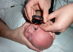
Methods: A retrospective observational case series. We reviewed and analyzed clinical records of all the premature infants born before the 32nd gestational week examined by ophthalmologist in ROP screening at the University Hospital in Ostrava in the period from 1. 9. 2011 to 31. 8. 2012. Children’s gestational age at birth, birth weight, postconceptional age (PCA) of the child at the time of the first ocular inspection, at the time of diagnosis ROP and at the time of any intervention, possible risk factors of ROP (Apgar score in the 1st minute, duration of oxygen therapy, FiO2 (%) (percentage fraction of oxygen in the inspired gas mixture), duration of mechanical ventilation, transfusion of erythrocytes (resuspended leukodepleted), presence of sepsis / infection in the perinatal period and duration of phototherapy) were evaluated. Eye examination was performed in local anesthesia with the use of an eyelid retractor, in artificial mydriasis, using an indirect ophthalmoscope and digital imaging system RetCam 3.
Results: 138 premature infants with an average gestational age at birth of 29.8 weeks, average birth weight 1385 g, were included in this study. Thirty-four children (24.6 %) were diagnosed with ROP, in all cases 1st stage at the time of diagnosis. An ophthalmologist indicated and subsequently implemented intervention (cryotherapy / laser treatment) in the case of five children (14.7 %) with ROP under general anesthesia. Average duration of oxygen therapy at infants with ROP was 371 hours, in the group without ROP 84 hours. The difference between the average values was statistically significant [t (37) = -3.69, P <= 0.0007]. Average time of mechanical ventilation in the case of children with ROP were 229 hours, in the group without ROP 41 hours [t (35) = -2.99, P <0.005]. In the case of children with ROP, we noticed on average 3 transfusions of erythrocytes, in the group without ROP 1 transfusion [t (40) = -3.94, P <= 0.0003]. The average value of the Apgar score in the 1st minute of children with ROP group was 6.3 and children without ROP 7.8. The difference between the average values of Apgar score in the 1st minute was between both groups statistically significant [t (136) = 4.06, P <= 0.00008]. Sepsis / infection in the perinatal period occurred in 30 (88.2 %) children with ROP, in comparison with 46 (44.2 %) children with sepsis / infection without ROP. Average duration of phototherapy in infants with ROP was 42.4 hours, in the group without ROP 53.6 hours [t(136) = 1,21, P<= 0,2]. Conclusion: This study demonstrated statistically significant correlation of Apgar score in the 1st minute, duration of oxygen therapy, duration of mechanical ventilation, transfusion of erythrocytes and presence of sepsis / infection on the onset and progression of ROP at premature infants in our group. No effect of FiO2 (%) and duration of phototherapy on the onset and progression of ROP was demonstrated.

References

  1. Akter, S., Hossain, M. M., Shirin, M. et al.: Blood Transfusion: A Risk Factor in etinopathy of Prematurity. Bangladesh J Child Hlth, 2012; 34, 2.
  2. Brooks, S.E., Marcus, D.M., Gillis, D., et al.: The Effect of Blood Transfusion Protocol on Retinopathy of Prematurity: A Prospective, Randomized Study. Pediatrics, 1999; 104: 3, 514–518.
  3. The system enables us to obtain photo documentation or a video recording from the examination, comparison of images in time and observation of the dynamics of progression of the disorder or effect of applicable treatment, the possibility of connection to the computer network, sharing and consultation of the obtained photo documentation with another workplace, in particular for borderline cases, and last but not least provides credible, defensible medical-legal documentation. Since December 2011 we have been using this equipment also for therapeutic interventions (laser photocoagulation/cryotherapy of the retina), and so far we have not encountered similar utilisation of this system in the literature. The obtained data shall be the subject of further analysis. CONCLUSION In our retrospective trial, in addition to the low birth weight and short gestation period of prematurely born infants, we identified also the length of oxygen therapy and the duration of artificial pul-
  4. Ebrahim, M., Ahmad, R., S., Mohammad, M.: Incidence and Risk Factors of Retinopathy of Prematurity in Babol, North of Iran. Ophthalmic Epidemiology. 2010; 17, 3: 166–170.
  5. Hakeem, A. H. A. A., Othman, M. F., Gamal, B. M.: Retinopathy of prematurity: A study of prevalence and risk factors. Middle East African Journal of Ophthalmology. 2012; 19, 3: 289.
  6. Health, P.: Pathology of retinopathy of prematurity, RLF. Am J Ophthalmol, 1953; 34: 1249–1259.
  7. Hungi, B., Vinekar, A., Datti, N., et al.: Retinopathy of Prematurity in a Rural Neonatal Intensive Care Unit in South India – A Prospective Study. The Indian Journal of Pediatrics, 2012; 79, 7: 911–915.
  8. Lorena, S. H. T., Brito, J. M. S.: Retrospec tive study of preterm newborn infants at the ambulatory of specialities Jardim Peri-Peri. Arquivos Brasileiros de Oftalmologia. 2009, roã. 72, ã. 3, s. 360-364. ISSN 0004-2749.
  9. Marková, A., Jurčuková, M., Dort, J. a kol.: Hodnocení rizikových faktorů vzniku ROP, oční vady a psychomotorický vývoj nedonošených dětí v západočeském regionu, dvanáctileté sledování. Čes a Slov Oftalmol, 2009; 65, 1: 24–28.
  10. Mehmet, S., Fusun, A., Sebnem, C., et al.: One-year experience in the retinopathy of prematurity: frequency and risk factors, short-term results and follow-up. Int J Ophthalmol, 2011; 4(6): 634-640.
  11. Mutlu, F., M., Altinsoy, H. I., Mumcuoglu, T., et al.: Screening for Retinopathy of Prematurity in a Tertiary Care Newborn Unit in Turkey: Frequency, Outcomes, and Risk Factor Analysis. J Pediatric Ophthalmol; 2008; 45, 5, 291–298.
  12. Odehnal, M., Malec, J., ·tûpánková, J., Dotfielová, D.: Souãasn˘ pohled na retinopatii nedono‰en˘ch. âes a Slov Oftalmol, 2011; 67, 2: 35–41.
  13. Palmert, E.A., Flynn, I.T., Hardy, R. J., et al.: for the Cryoteraphy for Retinopathy of prematurity Cooperative Group. Incidence and early course of retinopathy of prematurity. Ophthalmology, 1991; 98: 1628–1640.
  14. Reynolds, J. D., Hardy, R. J., Kennedy, K. A., et al.: Lack of Efficacy of Light Reduction in Preventing Retinopathy of Prematurity. New England J Med, 1998; 338, 22: 1572–1576.
  15. Saugstad, O.D.: Oxygen and retinopathy of prematurity. J Perinatol. 2006; 26 (suppl 1): 546–550.
  16. Schumann, R. de F., Barbosa, A. D. M., Valete, C. O.: Incidence and severity of retinopathy of prematurity and its association with morbidity and treatments instituted at Hospital Antonio Pedro from Universidade Federal Fluminense, between 2003 and 2005. Arquivos Brasileiros de Oftalmologia. 2010; 73, 1: 47–51.
  17. Terry, T. L.: Extreme Prematuruty and Fibroblastic Overgrowth of Persistent Vascular Shealth Behind Each Crystalline lens. I. Preliminary report. Am J Ophthalmol, 1942; 25: 203–204.
  18. The Incidence and Course of Retinopathy of Prematurity: Findings from the Early Treatment for Retinopathy of Prematurity Study. PEDIATRICS. 2005; 116, 1: 15–23.
  19. The photographic screening FOR retinopathy of prematurity study (PHO- TOROP). Retina. 2008; 28: Suppl, S47–S54.