
2014 - 2 Issue

Case report

Acute Posterior Multifocal Placoid Pigment Epitheliopathy – Case Report

Full Text links

Summary

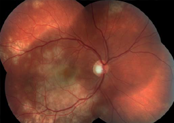
Acute posterior multifocal placoid pigment epitheliopathy (APMPPE) affects individuals between 20 to 30 years of age and ocular manifestations are often preceded by flu-like illness. Symptoms of the disease include acute vision decrease associated with central and paracentral scotoma. Impairment of vision is usually bilateral, but may be asymmetric. We report the case of a man at the age of twenty-one, who was treated for APMPPE. Due to the involvement of the macula, low visual acuity and related immunogenetic predisposition (HLA-B27 positivity) was recommended corticosteroid therapy with a good therapeutic effect.

References

  1. within the framework of differential diagnostics [11]. The majority of authors do not recommend the commencement of therapy of APMPPE, but in the case of the presence of negative prognostic factors, which are affliction of the macula, low visual acuity, connected immunogenetic predisposition, age higher than 60 years upon the first attack, unilateral manifestation of the disease and an interval of longer than 6 months before the affliction of the other eye, use of corticosteroids is recommended [3, 10]. In our case, with regard to the affliction of the central region, low visual acuity of the right eye and connected immunogenetic predisposition (HLA-B 27 positivity), general corticosteroid therapy was recommended, with good therapeutic effect. CONCLUSION Acute posterior multifocal placoid pigment epitheliopathy is an acquired inflammatory disease, which belongs to the heterogeneous group of white dot syndromes. In our case, on the basis of the clinical picture and anamnesis, we diagnos
  2. Allee, SD., Marks, SJ.: Acute posteri- or multifocal placoid pigment epitheli- opathy with bilateral central retinal vein occlusion, Am J Ophthalmol, 1998; 126: 309–312.
  3. Burés-Jelstrup, A., Adán, A., Casaroli-Marano, R.: Epiteliopatía pigmentaria placoide posterior multifocal aguda. Estudio de 16 casos., Arch Soc Esp Oftalmol, 2007; 82: 291-298
  4. De Souza, S., Aslanides, IM., Altomare, F.: Acute posterior multifocal placoid pigment epitheliopathy associated with retinal vasculitis, neovascularization and subhyaloid hemorrhage., Can J Ophthalmol, 1999; 34: 343–345.
  5. Ducos de Lahitte, G., Fajnkuchen, F., Giraud, C., Chabne, G.: Épithéliopathie en plaque et mauvais pronostic visuel: à propos d’un cas., J Fr Ophtalmol., 2004, 27: 617-622.
  6. Gass, JDM.: Acute posterior multifocal pigment epitheliopathy., Arch Ophthalmol., 1968; 80: 177–185.
  7. Gass, JDM.: Stereoscopic atlas of macular diseases: diagnosis and treatment., 3rd ed. St Louis: CV Mosby, 1987; pp. 504 –506.
  8. Holt, WS., Regan, CDJ., Trempe, C.: Acute posterior multifocal placoid pigment epitheliopathy. Am J Ophthalmol, 1976; 81: 403–412.
  9. Isashiki, M., Koide, H., Yamashita, T., Ohba, N.: Acute posterior multifocal placoid pigment epitheliopathy associated with diffuse retinal vasculitis and late haemorrhagic macular detachment., Br J Ophthalmol, 1986; 70: 255–259.
  10. Pagliarini, S., Piruet, B., Ffytche, TJ., Bird, AC.: Foveal involvement and lack of visual recovery in APMPPE associated with uncommon features., Eye, 1995, 9:42–47.
  11. Quillen, D., Davis, J., Gottlieb, J. et al.: The White Dot Syndromes., Am J Ophthalmol, 2004; 137:5 38–50
  12. Rose, SJ., Lou, PL.: Acute posterior multifocal placoid pigment epitheliopathy., in Albert, DM., Miller, JW.,: Principles and practice of ophtalmology II. Philadelphia, Elsevier; 2008., pp. 2089–2095.